Ludwig’s Angina

Early addressing and proper management with appropriate treatment planning could be a life-saving procedure

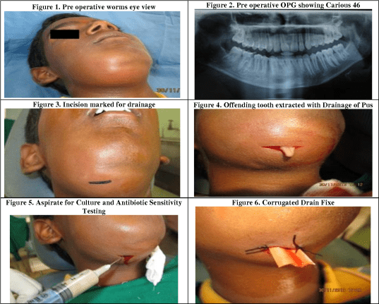
Ludwig’s angina is a form of severe diffuse cellulitis that occurs after a persistent odontogenic infection. Unless treated at an earlier stage, it could result in life-threatening situations.

It presents an acute onset and spreads rapidly, bilaterally affecting the submandibular, sublingual, and submental spaces, resulting in a state of emergency. The condition is relatively uncommon. Yet, it is potentially life-threatening due to the risk of impending airway obstruction. Taking into account its invasive nature and aggressiveness, early identification and management of Ludwig’s angina are extremely important.  Early addressing and proper management with appropriate treatment planning could be a life-saving procedure.

Ludwig’s Angina?

Ludwig’s angina was coined in 1836 after the German physician Wilhelm Frederick von Ludwig, who described this condition as a rapidly proliferating and progressive gangrenous cellulitis and swelling of the tissues of the skin in the face and neck region.

Ludwig’s Angina is considered fatal due to the nature of the swelling and the rapid spread of infection up to the neck, leading to a wide variety of complications, including airway obstruction, making it more lethal. Infection of the lower molars is the hallmark cause of true Ludwig’s angina, though this term is frequently applied to any floor of the mouth infection with sublingual and/or submaxillary space involvement.

How can this happen?

Ludwig’s angina usually originates as a dental infection of the second or third mandibular molars, including partially erupted third molars. The infection spreads lingually rather than buccally because the lingual aspect of the tooth socket is thinner. The infection initially spreads to the sublingual space and progresses to the submandibular space. The infection is usually polymicrobial involving oral flora, both aerobes and anaerobes. The most common organisms are Staphylococcus, Streptococcus, Pepto streptococcus, Fusobacterium, Bacteroides, and Actinomyces.  Immunocompromised patients are at a higher risk of Ludwig’s angina.

Ludwig’s angina and the dentist? How are they related?

There are a wide variety of causes for this condition, mainly including a decayed tooth, a space infection, a fracture of the lower jaw, an abscess pertaining to the lower jaw region, etc. Other causes include peritonsillar or parapharyngeal abscesses, mandibular fractures, oral lacerations/piercing or submandibular sialadenitis, and oral malignancy.

Preexisting factors include dental caries; recent dental treatment; systemic illnesses like diabetes mellitus, malnutrition, and alcoholism; compromised immune system such as AIDS; and organ transplantation.

How we manage it?

How we manage it1

FMS Dental Hospitals has an in-house operation theatre and a 24/7 monitoring intensive care unit with fully assisted experienced staff to manage the situation and provide adequate support to the patient.

As the size of the swelling is considerably large, patients will be dehydrated and compromised due to the inability to chew or swallow. To prevent this, we at FMS will administer adequate diet and nutritional support to the patient intravenously. The patient will be monitored and all the vital signs assessed by experienced staff to ensure a speedy recovery.

The experienced team at FMS, including anesthetists and oral and maxillofacial surgeons, will manage the airway obstruction, which is critical, followed by administration of IV, antibiotics, and analgesics to reduce the micro bacterial load in the area. For patients who don’t respond well to the initial antibiotics or in the case of a sizeable swelling, care needs to be given first to reduce the swelling and its associated contents. This is done by performing incision and drainage to reduce the pressure/infectious content inside the swelling. Corrugated rubber drains will be placed at multiple sites to reduce the pressure within the swelling and to prevent further spread, as any failure to intervene immediately can lead to a lot of complications including death.

After maintaining the vital signs, the foci of infection will be addressed. The cause should be removed at the earliest to prevent the further spread of infections. Dental extraction needs to be done after the primary management to eliminate the source of infection.

Tips to avoid this lethal condition

  • Proper maintenance of oral hygiene.
  • Stay within the limits of the underlying medical conditions as there may be a predisposing factor to worsen the condition.
  • Any concern should be addressed immediately. Early intervention can ensure better recovery.
  • Never delay any dental conditions or queries. We are here, just a call away.
  • Proper knowledge of the situation is a must for good treatment. Expert opinions should always be considered.
  • Faulty hands can easily worsen the situation. Make sure you trust yourself only with the right hands.

Cavernous Sinus Thrombosis
What is cavernous sinus thrombosis?

Cavernous sinus thrombosis (CST) is a rare, life-threatening disorder that can complicate facial infection, sinusitis, orbital cellulitis, pharyngitis, or otitis or occur following traumatic injury or surgery, especially in the setting of a thrombophilic disorder.

Early recognition of cavernous sinus thrombosis which often presents itself with fever, headache, eye findings such as periorbital swelling, and ophthalmoplegia, is critical for a good outcome.

The exact risks will differ for every person and some may be more specific to your case. This is why the initial consultation is so important, your surgeon will be able to assess the risks for you and advise you accordingly.

I am cavernous sinus thrombosis and this is how I present:

Cavernous sinus thrombosis is usually septic, but can also be aseptic. Septic cases can follow central facial infections, especially within the danger triangle of the face (from the corners of the mouth to the bridge of the nose). These include abscess or cellulitis, sinusitis (especially sphenoiditis and ethmoiditis), dental infections, extractions or procedures (even a posterior superior alveolar nerve block entering the pterygoid plexus), maxillofacial surgery, otitis media, and mastoiditis. Aseptic causes are less common than septic causes. These include trauma, surgery, or pregnancy.

Who is responsible?

Who is responsible

A variety of infectious organisms can cause cavernous sinus thrombosis although the majority are bacterial. Staphylococcus aureus may account for two-thirds of cases, and methicillin resistance should also be considered. Cavernous sinus thrombosis is so rare that incidence data is difficult to estimate. Since cavernous sinus thrombosis comprises approximately 1% to 4% of cerebral venous and sinus thrombosis (CVST), which has an annual incidence of approximately two to four per million people per year with a higher incidence in children, one can estimate that the annual incidence of cavernous sinus thrombosis might be approximately 0.2 to 1.6 per 100,000 per year. A male or female predominance in cavernous sinus thrombosis is uncertain.

The cavernous sinus has been called the “anatomic jewel box” because it shares an intimate relationship with several important structures. This is because a majority of vital anatomical structures pass through or near the cavernous sinuses.

How can we manage it?

Because of the rarity of diagnosis, no randomized controlled trials are available, and expert opinion guides treatment. In general, antimicrobial and antithrombotic therapies are primary considerations. No surgical interventions are recommended for the cavernous sinuses themselves. However, some patients might require sphenoidotomy, ethmoidectomy, maxillary antrostomy, mastoidectomy, abscess drainage, craniotomy (subdural empyema), orbital decompression, or ventricular shunt placement. Proper follow-up of patients is important even after the discontinuation of antibiotics.

Most Awarded Dental Clinic

phone

040 2222 1111

BOOK NOW

Experts Are Here to Help You!!