Root canal therapy has experienced a profound evolution in both perception and performance. Once regarded with apprehension due to discomfort and unpredictability, has become a predictable and comfortable intervention, supported by advanced technology, enhanced visualization, and improved clinical protocols. Today, modern endodontic therapy offers patients not only relief, but confidence in a treatment grounded in accuracy, efficiency, and long-term success.
One of the most important advancements in recent decades is Microscopic Root Canal Treatment also known as microscopic endodontics. While it may sound complex, what it really means for patients is better accuracy and better outcomes. This advanced technique uses a powerful dental operating microscope to help dentists see the inside of your tooth in extraordinary detail, far beyond what the naked eye allows. This enhanced visualization leads to more accurate diagnosis, more conservative treatment and improved long-term outcomes, particularly in complex cases.
At FMS Dental, successful root canal outcomes are driven by the seamless integration of advanced technology and exceptional clinical expertise. While the operating microscope significantly enhances precision and visibility, it is the skill, judgment, and experience of our endodontic specialists, Dr. Shekar, Dr. Priyendu Dr. Ravi Chandra, Dr. Naresh, Dr. Aisha Habeeb, Dr. Sandeep Eleti and other senior endodontists that truly ensure outstanding results.
Our team of endodontists (Root Canal Specialists) are extensively trained in root canal anatomy, microscopic techniques and the management of complex cases. Combined with state-of-the-art visualization technology, this expertise allows for treatments that are safer, more precise and consistently predictable.
At FMS Dental, this powerful synergy of innovation and experience ensures every patient receives care that is not only highly effective, but also comfortable and reassuring.
Understanding the Complexity Inside a Tooth
A tooth may seem simple and solid at first glance, yet beneath its surface lies a sophisticated living structure. Internally it contains a highly intricate system of canals that house the pulp (nerves, blood vessels, and connective tissue) making it vulnerable to infection or damage that often requires procedures like root canals to preserve its function.
These canals are often:
- Extremely narrow
- Curved or branching
- Partially or fully calcified
- Connected by microscopic isthmuses/tiny connecting passages
- Prone to cracks or structural defects
Even minor anatomical variations can influence treatment success. When parts of the canal system are not identified or adequately cleaned, infection can persist and lead to retreatment.
This is where magnification becomes crucial.
What Is Microscopic Root Canal Treatment?
Endodontic (root canal) treatment is a very precise and detailed procedure that requires a high level of focus and skill. The inside of a tooth is small and naturally dark, which can make it challenging to see clearly. Root canals can be extremely thin, sometimes even thinner than a strand of hair — and they may curve, branch out, or have tiny connecting pathways. Because of this complexity, careful attention to detail is essential to ensure the treatment is done thoroughly and accurately.
Microscopic root canal treatment is done using a special high-powered dental microscope. This microscope gives your dentist a much clearer, brighter, and magnified view of your tooth. With this enhanced visibility, your dentist can treat complex problems more precisely, including cases that might have been considered too difficult to treat in the past or that may not have had a good long-term outlook.
How Microscopic Technology Improves Your Root Canal Treatment
Modern root canal treatment has been transformed by advanced dental microscope technology, allowing for a higher level of precision and care than ever before. This technology gives your dentist a clear, magnified view of the inside of your tooth, making it easier to spot even the smallest details. With better visibility, treatment can be carried out more carefully and thoroughly, helping to improve results and protect your tooth. The table below shows how this technology makes your care safer, more precise, and more effective.
Features and Advancements
| Feature | What This Means for You |
| High magnification (3x–30x) | Your dentist can see your tooth in much greater detail than with the naked eye. |
| Focused, bright illumination | The inside of your tooth is clearly lit for better visibility during treatment. |
| Enhanced depth perception | Your dentist can better judge the exact position and depth of tiny structures inside the tooth. |
| Clear visualization of small details | Even very small or hidden canals can be located and treated properly. |
Impact on Visualization and Precision
| Improvement | What This Means for You |
| Enhanced visibility of root canal details | The inner structure of your tooth can be examined more thoroughly and accurately. |
| Precise identification of anatomical structures | Curved, narrow, or complex canals can be found and treated with greater precision. |
| Reduced procedural errors | Better visibility helps lower the risk of complications during treatment. |
Advantages Over Traditional Methods
| Advantage | What This Means for You |
| Better identification and treatment of complex canals | Teeth that may have been difficult to treat in the past can often be successfully treated. |
| Higher success rate | The improved precision increases the likelihood of a successful and long-lasting result |
Additionally, microscopes can record images, which greatly aids in doctor-patient communication.
For patients, this means a more thorough treatment, a lower chance of complications, and a reduced risk of needing retreatment in the future. Simply put, when your dentist can see better, they can treat better.
When Is Microscopic Root Canal Treatment Significantly Beneficial?
While most root canal procedures benefit from magnification, microscopic root canal treatment becomes especially valuable in complex or high-risk cases, where accuracy makes a real difference to long-term success.
Below are the clinical situations where microscopic treatment offers a substantial advantage:
1. When a Previous Root Canal Has Failed
If a tooth needs retreatment, the reason is often hidden deep within the canal system—such as a missed canal or lingering infection. The microscope helps your dentist identify and correct these issues with greater precision, improving the chances of saving the tooth.
2. In Calcified or Very Narrow Canals
Over time, canals can become extremely narrow due to aging or past trauma. Magnification allows careful location and treatment of these canals while preserving healthy tooth structure.
3. If an Instrument Breaks Inside the Canal
On rare occasions, a small file may fracture during treatment. Microscopic visualization makes safe removal more predictable and conservative.
4. When a Crack or Fracture Is Suspected
Tiny cracks can be difficult to detect. The microscope helps confirm an accurate diagnosis and guide the right treatment plan.
5. During Endodontic Surgery (Apicoectomy)/ Root end surgery
In surgical procedures, magnification enhances precision, leading to better healing and higher success rates.
6. For Front Teeth Where Aesthetics Matter
Microscopic access allows smaller, more conservative openings—helping preserve strength and appearance.
While not every root canal requires advanced magnification, in technically demanding situations, the microscope transforms treatment from estimation to precision-guided care, significantly improving predictability and outcomes
Enhancing Root Canal Precision with Advanced Microscopes at FMS Dental
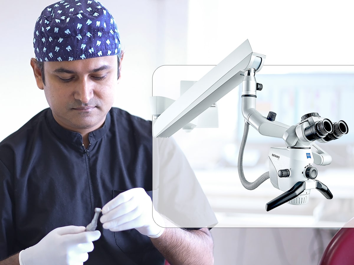
At FMS Dental, clinical excellence is supported by world-class visualization systems. The use of advanced dental operating microscopes to perform microscopic root canal treatment, a gold-standard approach in modern endodontics that significantly improves precision, visibility, and treatment outcomes. These microscopes provide high magnification, shadow-free illumination, ergonomic control, and optional imaging capabilities, allowing our clinicians to locate and treat even the smallest and most complex root canal anatomy.
Seiler Dental Microscopes
Seiler systems are globally recognized in endodontic microsurgery and root canal therapy for their optical quality and ergonomic design. They enable precise identification of canal orifices, tiny fractures, and anatomical variations which are critical for success in root canal disinfection and filling.
Seiler Alpha Air Series and the Seiler 3D Dental Surgical Microscope, these state-of-the-art dental operating microscopes combine multi-level magnification, premium German Schott glass optics, and coaxial LED illumination to deliver exceptional clarity and shadow-free visualization of intricate root canal anatomy. With high-definition 3D imaging and magnification capabilities of up to 28×, they significantly enhance diagnostic precision, clinical control, and ergonomic efficiency, ensuring predictable, highly refined outcomes in even the most complex endodontic procedures.
Labomed Dental Operating Microscopes
Labomed Dental Operating Microscopes are also part of the advanced diagnostic and treatment setup at FMS Dental:
The Labomed Magna delivers superior magnification and crystal-clear stereo optics for identifying complex root anatomy, while the Labomed Prima DNT features smooth motorised focusing for precise, ergonomic adjustments during treatment.
These Labomed microscopes deliver high-contrast, deep-field optical performance, which is essential for locating hidden canals and ensuring thorough cleaning and shaping of the root canal system.
How It Helps Patients at FMS Dental
Using these microscopes, the endodontic specialists can:
- Locate and treat even the smallest and most complex canals
- Detect cracks, fractures, or hidden anatomy with confidence
- Reduce procedural errors and improve healing outcomes
- Perform conservative preparation with minimal tooth removal
This translates into higher success rates, fewer complications, and more predictable long-term results in root canal therapy.
Expert Insights from the Endodontic Team at FMS
Technology has revolutionized root canal treatment, making it safer, more accurate, and more comfortable than ever before. Microscopic endodontics represents a major advancement in the way root canal therapy is performed. It is not a cure-all for every dental condition, but it significantly elevates diagnostic accuracy, procedural control, and long-term outcomes, especially in complex or retreatment cases.
At FMS Dental, our investment in advanced dental technology reflects our commitment to delivering exceptional care and long-lasting results. By combining innovation with expert clinical skill, we ensure that every root canal treatment is performed with precision, confidence, and care, helping our patients preserve their natural smiles for years to come.
If you are facing root canal therapy or retreatment, discussing microscopic endodontic options with your clinician may help ensure the highest level of precision and care.
Consult FMS Dental for Advanced Root Canal Treatments. Schedule your consultation today !!
For Appointment booking. Please call us on 8885060770 or 04022221111 or email us at [email protected]
Frequently Asked Questions About Microscopic Root Canal Treatment
Here are some commonly asked questions about microscopic root canal treatment to help you better understand the procedure and its benefits.
What is Microscopic Root Canal Treatment?
Microscopic Root Canal Treatment is an advanced endodontic procedure performed using a high-magnification dental operating microscope. This technology provides superior illumination and clarity, allowing the endodontist to see intricate internal tooth structures that are not visible to the naked eye. It enables precise cleaning, shaping, and sealing of the root canal system. The result is a more accurate and predictable treatment outcome.
How does a dental microscope improve root canal treatment?
A dental microscope significantly enhances visibility inside the tooth. It helps detect hidden canals, micro-fractures, calcifications, and complex anatomy that could otherwise be missed. With magnification and focused lighting, the endodontist can perform each step with greater control and precision. This reduces procedural errors and improves the overall quality of treatment.
Is microscopic root canal treatment more successful than conventional treatment?
Yes, microscope-assisted treatment is associated with higher success rates, particularly in complex cases. The enhanced visualization reduces the chances of missed canals — one of the main causes of root canal failure. It also allows for more thorough cleaning and better sealing of the canals. This improves long-term tooth survival and reduces the likelihood of retreatment.
Is the microscopic root canal procedure painful?
Microscopic root canal treatment is performed under local anaesthesia, just like a conventional root canal. Patients typically experience minimal discomfort during the procedure. Because the treatment is more precise and less invasive to surrounding tissues, post-treatment recovery is often smoother. Most patients return to normal activities shortly afterward.
When is microscopic root canal treatment especially recommended?
It is particularly recommended for teeth with complex or curved canals, calcified canals, or previously failed root canals requiring retreatment. It is also valuable in cases where cracks or unusual anatomy are suspected. In such situations, magnification greatly improves diagnostic accuracy and treatment precision. This increases the chances of saving the natural tooth.
Can microscopic treatment help save severely damaged teeth?
Yes, in many cases it can. The microscope allows the clinician to detect fine fractures, accessory canals, and hidden infections that might otherwise go unnoticed. Early identification and precise management improve the likelihood of preserving even structurally compromised teeth. Preserving natural dentition is always the primary goal of endodontic care.
Does using a microscope make the procedure longer?
Not necessarily. While highly complex cases may require additional attention, improved visualization often makes treatment more efficient. The ability to clearly see the canal anatomy reduces guesswork and repeated attempts. In many cases, this efficiency actually saves time and reduces the need for future corrective procedures.
Is microscopic root canal treatment safe?
Yes, it is a safe and evidence-based advancement in endodontics. The technology enhances precision without increasing procedural risk. In fact, better visibility helps minimize complications such as perforations or missed anatomy. It supports a more conservative approach that preserves healthy tooth structure.
Does every root canal require a microscope?
Not every case strictly requires one, but using a microscope significantly improves treatment quality in most situations. Many modern endodontic practices consider it the standard of care, especially for molars and complex anatomy. Even in routine cases, magnification enhances accuracy and attention to detail. It represents a higher level of clinical precision.
Is microscopic root canal treatment more expensive?
Because it involves advanced technology and specialized expertise, the cost may be slightly higher than conventional treatment. However, the improved accuracy and long-term predictability often make it a worthwhile investment. Reducing the risk of retreatment ultimately saves time, cost, and discomfort in the future. The focus is on durable, high-quality results.
References
1. Effectiveness of microscope-assisted root canal treatment in permanent posterior teeth: A retrospective cohort study. Journal of Dentistry, 157, 105771.
2.The clinical treatment of complicated root canal therapy with the aid of a dental operating microscope. International dental journal, 61(5), 261-266.
3. Comparative Analysis of Latest Technologies in Microscopic Endodontics: Diagnostic. Journal of Pioneering Medical Sciences, 13, 128-137.